What is the spinal cord?
The spinal cord is part of the nervous system and is about 45 cm long in men and 43 cm long in women. The length of the spinal cord is much shorter than the length of the bony spinal column. It runs the length of the back, extending from the base of the brain to about the waist. The area within the vertebral column beyond the end of the spinal cord is called the cauda equina. The nerves that branch out from the spinal cord to the other parts of the body are called lower motor neurons (LMNs) and dorsal root sensory neurons. These spinal nerves exit and enter at each vertebral level and communicate with specific areas of the body.
 What is a spinal cord injury? What is a spinal cord injury?
Spinal cord injury (SCI) is damage to the nerves within the spinal canal, thereby affecting the spinal cord's ability to send and receive messages from the brain to the body's systems that control sensory, motor and autonomic function below the level of injury. A SCI can be complete or incomplete. In a complete injury, nerve damage obstructs every signal coming from the brain to the body parts below the injury. In an incomplete injury, some residual motor and sensory function remains below the level of SCI.
The self healing capability of the body can repair some of the spinal cord injury, but surely not all. I used to have a complete injury, but now 11 years after the accident I can feel again for about 25%, and also do a couple of muscles work again. The body needs a lot of help to fully recover from a spinal cord injury. The spinal cord is about the most complex body part. To help the body repair it 100%, a lot of research and development needs to be done, which clearly is no priority of any government I know. When if the spinal cord can be repaired, just about any other injury could be repaired.
A spinal cord injury compared to a broken machine
Obviously, a biological nervous system isn't equal to an electronical or mechanical machine, but there are some similarities. A computer processor chip is basically an awful amount of simple switches, and a computer memory chip is simply a huge collection of buckets that do or do not contain electrons. It's not the quality of bits that make good software. What matters is the structure, the arrangement of bits.
One nerve is complex relative to one electro/mechanical switch, it's like a small chemical factory that's alive. But then again is its function pretty simple. There are just a few types, but each neuron is unique in it's place and connections, and they are present in huge amounts, billions, arranged in such fashion that they together are the nervous system of a human body. The difference between people's nervous system (including the brain) is not really the kind or amount of nerves, but the unique structure.
I have repaired some mechanical photo cameras. When if a camera is broke, opening it, poring oil in it and closing it, will NOT repair it. This is why I don't believe that simply injecting stem-cells will repair an injured spinal cord. To repair a mechanical photo camera:
- Open the thing, without causing damage.
- Take it completely apart, if necessary.
- Clean all parts, thoroughly.
- Replace broken parts, if necessary. Most often, dirt is the only problem!
- Put it together again, use the right kind of lubricant where needed.
- Try all functions many times, adjust pressures if necessary.
- Close it nicely, tadaa; ready.
For most repairs it's not necessary to fully understand every part of the system.
What similarities are there between a broken spinal cord (a biological system) and a broken camera (a electro-mechanical system)?
Can this bodypart be replaced by a machine-like part, or should it be regenerated completely biological, also to prevent a rejection from the body?
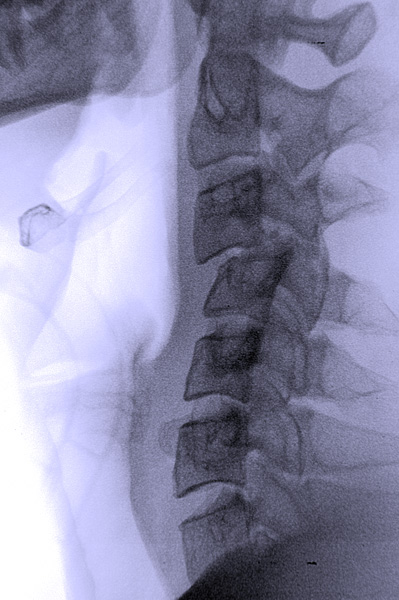
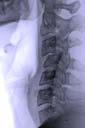
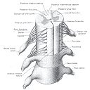
Anatomy of the Vertebrae
 The rings of bone that make up the spinal column are known as vertebrae. The vertebrae are named according to their location on the spinal column and are called the: Cervical, Thoracic, Lumbar and Sacral vertebrae. The spinal cord is located in the vertebral foramen and is made up of 31 segments: 8 cervical, 12 thoracic, 5 lumbar, 5 sacral and 1 coccygeal. A pair of spinal nerves exits from each segment of the spinal cord. The rings of bone that make up the spinal column are known as vertebrae. The vertebrae are named according to their location on the spinal column and are called the: Cervical, Thoracic, Lumbar and Sacral vertebrae. The spinal cord is located in the vertebral foramen and is made up of 31 segments: 8 cervical, 12 thoracic, 5 lumbar, 5 sacral and 1 coccygeal. A pair of spinal nerves exits from each segment of the spinal cord.
The seven vertebrae in the neck are the Cervical Vertebrae. SCI at cervical levels usually causes a loss of independent breathing and loss of function to the arms and legs, thereby resulting in quadriplegia.
The twelve vertebrae in the chest are called the Thoracic Vertebrae. Thoracic level injuries usually affect the chest and the legs and result in paraplegia.
The five vertebrae in the lower back are known as the Lumbar Vertebrae. Lumbar level injury typically results in loss of control of the legs, bladder, bowel and sexual functions.
The Sacral Vertebrae are the five vertebrae that run from the pelvis to the end of the spinal column. Sacral level injuries generally damage the nerves emanating from the distal spinal cord conus and typically cause lower motor neuron flaccid paralysis type lesions involving some loss of function in the legs and difficulty with bowel, bladder and sexual control.
Neuron Structure
Nerve cells are called neurons. Neurons have the ability to gather and transmit electrochemical signals. Neurons share the same characteristics and have the same parts as other cells, but the electrochemical aspect lets them transmit signals over long distances (up to several a few meters) and pass messages to each other.
Neurons have three basic parts:
1) Cell body - This main part has all of the necessary components of the cell, such as the nucleus (contains DNA), endoplasmic reticulum and ribosomes (for building proteins) and mitochondria (for making energy). If the cell body dies, the neuron dies.
2) Axon - This long, cable-like projection of the cell carries the electrochemical message (nerve impulse or action potential) along the length of the cell.
Note: Depending upon the type of neuron, axons can be covered with a thin layer of myelin, like an insulated electrical wire. Myelin is made of fat, and it helps to speed transmission of a nerve impulse down a long axon. Myelinated neurons are typically found in the peripheral nerves (sensory and motor neurons), while non-myelinated neurons are found in the brain and spinal cord.
3) Dendrites or nerve endings - These small, branch-like projections of the cell make connections to other cells and allow the neuron to talk with other cells or perceive the environment. Dendrites can be located on one or both ends of the cell.
Neurons and Synapses
The basic computational unit in the nervous system is the nerve cell, or neuron. A neuron has:
* Dendrites (inputs) a neuron
* Cell body
* Axon (output)
A neuron receives input from other neurons (typically many thousands). Inputs sum (approximately). Once input exceeds a critical level, the neuron discharges a spike - an electrical pulse that travels from the body, down the axon, to the next neuron(s) (or other receptors). This spiking event is also called depolarization, and is followed by a refractory period, during which the neuron is unable to fire.
The axon endings (Output Zone) almost touch the dendrites or cell body of the next neuron. Transmission of an electrical signal from one neuron to the next is effected by neurotransmittors, chemicals which are released from the first neuron and which bind to receptors in the second. This link is called a synapse. The extent to which the signal from one neuron is passed on to the next depends on many factors, e.g. the amount of neurotransmittor available, the number and arrangement of receptors, amount of neurotransmittor reabsorbed, etc.
The human brain contains about 10 billion nerve cells, or neurons. On average, each neuron is connected to other neurons through about 10,000 synapses. (The actual figures vary greatly, depending on the local neuroanatomy)
Neurons typically operate at a maximum rate of about 100 Hz
Basic Neuron Types
Neurons come in many sizes. For example, a single sensory neuron from your fingertip has an axon that extends the length of your arm, while neurons within the brain may extend only a few millimeters. Neurons have different shapes depending on what they do. Motor neurons that control muscle contractions have a cell body on one end, a long axon in the middle and dendrites on the other end; sensory neurons have dendrites on both ends, connected by a long axon with a cell body in the middle.
Some types of neurons:
- Bipolar (Interneuron)
- Unipolar (Sensory Neuron)
- Multipolar (Motoneuron)
- Cortical Pyramidal Cell
Neurons also vary with respect to their functions:
* Sensory neurons carry signals from the outer parts of your body (periphery) into the central nervous system.
* Motor neurons (motoneurons) carry signals from the central nervous system to the outer parts (muscles, skin, glands) of your body.
* Receptors sense the environment (chemicals, light, sound, touch) and encode this information into electrochemical messages that are transmitted by sensory neurons.
* Interneurons connect various neurons within the brain and spinal cord.
Neuronal types
Motor neurons
These lower motor neurons are located on the ventral aspect of the cord. They are either alpha or gamma cells.
- Alpha cells are the principle lower motor neurons of the spinal cord and form the main portion of the final common pathway. They conduct rapid motor impulses, with each alpha cell innervating approximately 200 muscle fibers.
- Gamma neurons are also part of the final common pathway according to some sources but they are only half as numerous as alpha cells. Gamma cells conduct slow motor impulses. Their major function is to stretch muscle spindles.
Association neurons
Interneurons connect the anterior and posterior horns of the gray matter and are involved in the reflex arc. They work within the same segment of the spinal cord, with a segment being defined as the horizontal section of the cord that gives rise to one pair of spinal nerves.
Internuncial Neurons travel between segments, sending projections up to the brain stem and cerebellum. They project in an ascending, not descending manner.
These association neurons are found throughout the central nervous system. They are much more numerous than motor neurons; the ratio between the two types of cells is 30:1.
The main function of the association neurons in the spinal cord is that of inhibitory control. They also interconnect other cells with one another.
Some sources, including Bhatnager and Andy, (1995), do not distinguish between interneurons and internuncial neurons. Even if these two types of association neurons are grouped together, they should definitely be distinguished from the spinal nerves which are lower motor neurons, forming a final common pathway for information descending from the brain.
|
|
Major Divisions of the Brain
* Spinal Cord - The spinal cord can be viewed as a separate entity from the brain or merely as a downward extension of the brainstem. It contains sensory and motor pathways from the body, as well as ascending and descending pathways from the brain. It has reflex pathways that react independently of the brain, as in the knee-jerk reflex.
* Brainstem - The brainstem consists of the medulla (an enlarged portion of the upper spinal cord), pons and midbrain (lower animals have only a medulla). The brainstem controls the reflexes and automatic functions (heart rate, blood pressure), limb movements and visceral functions (digestion, urination).
* Cerebellum - The cerebellum integrates information from the vestibular system that indicates position and movement and uses this information to coordinate limb movements.
Forebrain: Diencephalon - thalamus, hypothalamus & Cerebral cortex
* Hypothalamus and pituitary gland - These control visceral functions, body temperature and behavioral responses such as feeding, drinking, sexual response, aggression and pleasure.
* Cerebrum (also called the cerebral cortex or just the cortex) - The cerebrum consists of the cortex, large fiber tracts (corpus callosum) and some deeper structures (basal ganglia, amygdala, hippocampus). It integrates information from all of the sense organs, initiates motor functions, controls emotions and holds memory and thought processes. The cerebrum is the largest part of the human brain.
In addition, the part of the brain called the thalamus evolved to help relay information from the brainstem and spinal cord to the cerebral cortex.
Lower Brain
The basic lower brain consists of the spinal cord, brainstem and diencephalon (the cerebellum and cortex are also present here..). Within each of these structures are centers of neuronal cell bodies, called nuclei, that are specialized for particular functions (breathing, heart-rate regulation, sleep):
* Medulla - The medulla contains nuclei for regulating blood pressure and breathing, as well as nuclei for relaying information from the sense organs that comes in from the cranial nerves.
* Pons - The pons contains nuclei that relay movement and position information from the cerebellum to the cortex. It also contains nuclei that are involved in breathing, taste and sleep.
* Midbrain - The midbrain contains nuclei that link the various sections of the brain involved in motor functions (cerebellum, basal ganglia, cerebral cortex), eye movements and auditory control. One portion, called the substantia nigra, is involved in voluntary movements; when it does not function, you have the tremored movements of Parkinson's disease.
* Thalamus - The thalamus relays incoming sensory pathways to appropriate areas of the cortex, determines which sensory information actually reaches consciousness and participates in motor-information exchange between the cerebellum, basal ganglia and cortex.
* Hypothalamus - The hypothalamus contains nuclei that control hormonal secretions from the pituitary gland. These centers govern sexual reproduction, eating, drinking, growth, and maternal behavior such as lactation (milk-production in mammals). The hypothalamus is also involved in almost all aspects of behavior, including your biological "clock," which is linked to the daily light-dark cycle (circadian rhythms).
Overview:
The Nervous System
Anatomical planes
The brain can be dissected for study in several ways:
- A medial cut or section divides the brain into right and left halves of equal size, separating the right and left hemispheres from one another.
- A sagittal cut runs parallel to the medial cut, but divides the brain into right and left portions of unequal size. A medial section may be considered to be a type of sagittal cut. However, a sagittal section is not a type of medial cut.
- A coronal cut runs from ear to ear, separating the brain into front and back portions.
- Horizontal (equal halves) or transverse cuts are perpendicular to coronal, medial, and sagittal cuts. They divide the brain into upper and lower sections.
When describing the nervous system, anatomists use the terms:
- Anterior and posterior to indicate front and back.
- Superior and inferior are used to refer to the upper and lower parts of the nervous system.
- Cranial and cephalic may be used as synonyms for superior.
- Rostral, which literally means "toward the beak," is also sometimes substituted for superior.
- The antonym for rostral is caudal, a term that means "toward the tail," and may be used to replace inferior in descriptions of the brain and spinal cord.
- Ventral means "toward the belly" and dorsal means "toward the back." Structures in the lower part of the brain may be described as ventral.
- Medial means toward the center while the term lateral signifies toward the sides.
Central Nervous System
The neuraxis or central nervous system consists of the brain and spinal cord.
The Brain is made up of the cerebral cortex , sub cortical structures, brain stem and cerebellum.
The spinal cord consists of grey and white matter surrounded by meninges in which cerebro-spinal fluid circulates. It runs from just below the medulla to small of back. Below that the cauda equina consisting of projections from the spinal cord goes down to the coccygeal area.
Peripheral Nervous System
Cranial Nerves:
There are twelve pairs of cranial nerves,
Ten of them have their cell bodies in the brain stem.
Some are motor; some are sensory, and some are both motor and sensory.
Six of them are involved in speech and swallowing.
Spinal Nerves:
They connect the central nervous system to the body.
There are thirty-one pairs each of which is both sensory and motor.
Autonomic Nervous System
Distribution:
Involved in control of all automatic and glandular functions, it is controlled by the hypothalamus.
It works with the the endochrine system for control of hormonal secretion.
Sympathetic and Parasympathetic Divisions:
Sympathetic prepares the body for flight or fight.
Parasympathetic helps, among other things, to bring the body back to normal.
Meninges and cerebrospinal fluid
The brain and spinal cord are covered by a series of tough membranes called meninges, which protect these organs from rubbing against the bones of the skull and spine. For further protection, the brain and spinal cord float in a sea of cerebrospinal fluid within the skull and spine. This cushioning fluid is produced by the choroid plexus tissue, which is located within the brain, and flows through a series of cavities (ventricles) out of the brain and down along the spinal cord. The cerebrospinal fluid is kept separate from the blood supply by the blood-brain barrier.
The Spinal Nerves
Description.
There are thirty-one pairs of spinal nerves. These nerves are mixed, having both a sensory and a motor aspect. Their motor fibers begin on the ventral part of the spinal cord at the anterior horns of the gray matter. The roots of their sensory fibers are located on the dorsal side of the spinal cord in the posterior root ganglia. When the motor and sensory fibers exit the spinal column through the intervertebral foramina and pass through the meninges, they join together to form the spinal nerves.
Spinal nerves receive only contralateral innervation from first order neurons.
Eight pairs of spinal nerves are located in the uppermost, cervical region of the cord:
- Twelve pairs are found in the thoracic region.
- Five pairs are in the lumbar area.
- Five pairs are in the sacral area.
- One pair is found in the most inferior, coccygeal region.
Function
These second order lower motor neurons, the spinal nerves, form part of the final common pathway for information traveling from the central nervous system to the periphery. The spinal nerves provide innervation to body areas below the neck while cranial nerves (also second order neurons) carry impulses only to the head and neck, except for the vagus. (You will understand shortly that cranial nerves can be sensory, motor or both).
Reflex arc
Also, the sensory and motor fibers of the spinal nerves form a reflex arc. This type of reflexive behavior occurs when a message from sensory fibers causes a motor reaction directly, without traveling to the brain. For example, if you touch a hot burner on the stove, sensory information about the temperature of the burner travels along spinal nerves to your spinal cord and are carried directly to their motor nuclei by interneurons; the motor command goes out along the axons of the lower motor neuron causing you to move your hand away from the stove. As messages do not have to travel up to the brain to be processed, reactions mediated by this reflex arc can occur very rapidly. Of course you WILL feel pain shortly thereafter (milliseconds) as the information gets to the parietal lobe via the thalamus
White and Gray matter
There is differences in the shape and size of the spinal cord at different levels. The dark gray color in each segment represents "gray matter." Nerve cell bodies are located in the gray matter. Surrounding the gray matter is white matter (lighter color shading) - this is where the axons of the spinal cord are located.
Compare the relative amount of gray and white matter at each level of the spinal cord. In the cervical segment, there is a relatively large amount of white matter. This pattern is caused by the many axons going up to the brain from all levels of the spinal cord AND there are many axons traveling from the brain down to different segments of the spinal cord. In lower segments of the spinal cord, there is less white matter because there are fewer axons traveling to and from the brain.
 Dorsal root ganglion Dorsal root ganglion
Receptors in the skin send information to the spinal cord through the spinal nerves. The cell bodies for these nerve fibers are located in the dorsal root ganglion. The nerve fibers enter the spinal cord through the dorsal root. Some fibers make synapses with other neurons in the dorsal horn, while others continue up to the brain. Many cell bodies in the ventral horn of the spinal cord send axons through the ventral root to muscles to control movement.
The Autonomic Nervous System
The autonomic nervous system is involved in the control of the heart, glands and smooth muscles of the body and plays a major role in regulating unconscious, vegetative functions. It works together with the endocrine system to control the secretion of hormones and is itself controlled by the hypothalamus.
Although the autonomic nervous system is considered to be one of the three main divisions of the human nervous system in its own right, parts of both the central nervous systems and the peripheral nervous systems play a role in its functions.
The autonomic nervous system has two components, the sympathetic system and the parasympathetic system. These two aspects have antagonistic functions.
The autonomic nervous system consists of four chains of nuclei or ganglia, two of which are located on either side of the spinal cord. The outer chains of nuclei form the parasympathetic division of the system while those closest to the spinal cord make up its sympathetic element.
|
|